Mechanical Engineering learning from biology and mastering biology: Biomechanics
It is well known that muscles become thicker and stronger with exercise, and they become thinner and weaker when not used. Additionally, exposure to microgravity in space causes calcium to leach from bones,
leading to a decrease in bone strength within just one week. Alternatively,
the walls of the heart and arteries exposed to high blood pressure become thicker compared to
those under normal blood pressure conditions(Figure 1).
Why do these changes occur?

It seems that biological tissues such as muscles, bones, heart, and blood vessels require "appropriate forces" to maintain healthy function. When these forces change, biological tissues can adapt to restore their normal function. So, what kinds of forces are necessary for the maintenance of biological function? How do biological tissues sense these forces? Furthermore, could leveraging the relationship between biology and forces lead to new treatments for diseases that were previously difficult to treat? The response to applied forces is not limited to macroscopic tissues alone but also occurs in the microscopic cells that make up tissues. For example, endothelial cells lining the inside of blood vessels (Figure 2) are known to be elongated and oriented in the direction of blood flow (Figure 3). Smooth muscle cells, which form the blood vessel wall itself, are understood to enlarge and thicken in response to applied forces. Moreover, when these cells are cultured attached to an elastic membrane and subjected to stretching and relaxation, they are known to orient themselves perpendicular to the direction of stretch.

 Endothelial cells aligning in the direction of blood flow is believed to be a response aimed at reducing the shear stress (the force that tries to peel cells away from the wall) exerted by the flow. Similarly, the enlargement of smooth muscle cells is thought to be a response aimed at maintaining a constant tensile stress (tensile force per unit cross-sectional area) on the cells themselves. Additionally, these cells align perpendicular to the direction of stretch to minimize the strain they experience due to stretch.
Similar examples are known for bones. Inside bones, thin column-like structures called trabeculae run extensively in all directions (Figure 4), and the orientation of these trabeculae is said to align with the principal stress directions (where tensile or compressive forces are greatest) (Figure 5). It has been noted since the 19th century (Wolff's Law) that the shape of bones itself achieves maximum strength with minimal material in response to applied forces.

 Furthermore, it is known that when an artery is cut like a ring and one section of the ring is incised, the ring opens up into an arc shape (Figure 6). This demonstrates that before cutting, there was residual compressive stress on the inner side of the ring and tensile stress on the outer side. It is hypothesized that this occurs because the inner and outer sides of the artery wall, under pressure from blood flow, bear equal amounts of tensile stress (Figure 7).

 In this way, biological tissues not only respond to forces but their responses are also believed to achieve optimal states in some sense. Thus, could we apply these biological properties to design mechanically optimal mechanical components? Or could we create sensors based on these principles? Moreover, is it possible to enable living organisms to produce mechanical components themselves?
The academic field that studies the mechanisms, structures, and behaviors of living organisms from a mechanical perspective is called Biomechanics. It involves observing and analyzing biological phenomena through the lens of essential mechanical engineering subjects such as materials mechanics, fluid mechanics, and mechanical dynamics. In our laboratory, we focus on biomechanics of cells and soft biological tissues, aiming to elucidate the relationship between these forces and biological processes using engineering methods. Furthermore, we strive to apply the knowledge gained to medicine and engineering, advancing our research day by day.

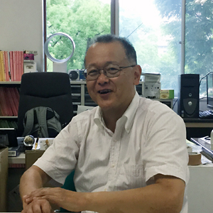
Prof.Takeo Matsumoto

Figure 1. Cross-sectional images of the thoracic aorta in normotensive (left) and hypertensive (right) rats.
The section of the thoracic aorta, parallel to the vessel axis, were stained with Azan-Mallory. The luminal side on the left. Red indicates smooth muscle cells, the white layer is the elastic lamina (elastin), and blue represents collagen fibers. Each specimen was fixed with formalin at the blood pressure of each rat. When hypertension occurs, the wall thickens, but the number of layers does not change. It is also evident that the innermost layer is particularly thickened. The inner side of the vessel wall is where the circumferential tensile stress increases due to the rise in internal pressure. In other words, it is inferred that the smooth muscle cells in the areas of higher stress are more hypertrophic (Matsumoto and Hayashi, 1996).It seems that biological tissues such as muscles, bones, heart, and blood vessels require "appropriate forces" to maintain healthy function. When these forces change, biological tissues can adapt to restore their normal function. So, what kinds of forces are necessary for the maintenance of biological function? How do biological tissues sense these forces? Furthermore, could leveraging the relationship between biology and forces lead to new treatments for diseases that were previously difficult to treat? The response to applied forces is not limited to macroscopic tissues alone but also occurs in the microscopic cells that make up tissues. For example, endothelial cells lining the inside of blood vessels (Figure 2) are known to be elongated and oriented in the direction of blood flow (Figure 3). Smooth muscle cells, which form the blood vessel wall itself, are understood to enlarge and thicken in response to applied forces. Moreover, when these cells are cultured attached to an elastic membrane and subjected to stretching and relaxation, they are known to orient themselves perpendicular to the direction of stretch.

Figure 2. Structure of an artery.
Blood vessel walls are divided into three layers: the intima, media, and adventitia, from the inside out. The intima consists of endothelial cells aligned along the vascular axis and the internal elastic lamina (mainly composed of elastin) to which these cells are attached. The media consists of smooth muscle cells, elastin, and collagen aligned circumferentially. The adventitia is composed of fibroblasts and collagen fibers. Strictly speaking, this diagram represents a muscular artery where the media is primarily composed of smooth muscle. Elastic arteries, such as the aorta and common carotid artery, have a slightly different structure. Specifically, in elastic arteries, the media has a structure where layers of smooth muscle and elastic lamina alternate, as shown in Figure 1. This repeating unit is called a lamellar unit (modified from “Atlas of the Body,” Kodansha, 1989).
Figure 3. Response of bovine endothelial cells to flow.
When endothelial cells isolated from the inner surface of the aorta are cultured in a petri dish, they adhere to the bottom and spread out in a cobblestone-like monolayer (right). When shear stress is applied to the cells by adding flow to the culture medium, the cells elongate into a spindle shape and align in the direction of the flow (left). The figure shows stained observations of actin filaments, one of the fine structures within the cells (Kataoka et al., 1997).Similar examples are known for bones. Inside bones, thin column-like structures called trabeculae run extensively in all directions (Figure 4), and the orientation of these trabeculae is said to align with the principal stress directions (where tensile or compressive forces are greatest) (Figure 5). It has been noted since the 19th century (Wolff's Law) that the shape of bones itself achieves maximum strength with minimal material in response to applied forces.

Figure 4. Cross-section of the proximal end of the femur.

Figure 5. Theoretical solution of the optimal shape of a beam under uniformly distributed load at its tip (left) and the similarity to the shape of the femur (right) (Thompson, 1942).
The curves inside the theoretical solution indicate the direction of principal stresses, while the curves inside the femur indicate the direction of the trabeculae. Not only are the shapes similar, but the direction of the trabeculae remarkably aligns with the direction of the principal stress lines.
Figure 6. Cross-sectional sample of a dog's thoracic aorta (left) and one that has been cut and opened into an arc.
The fact that the ring opens up when cut indicates that, despite the cross-sectional sample being in a no-load state (no external force acting), it was not stress-free. There were residual stresses: compressive stress on the inner wall and tensile stress on the outer wall.
Figure 7. Circumferential stress distribution in a blood vessel.
As shown in the top panel, if there were no residual stresses in the no-load state of the vessel, the circumferential stress in the loaded state (physiological state) would be higher on the inside and lower on the outside, as understood by considering the "deformation of a thick-walled cylinder " in the strength of materials. Conversely, as shown in the bottom panel, if the stress distribution were uniform across the vessel wall thickness in the physiological state, the no-load sample would have residual compressive stress on the inside and tensile stress on the outside. Cutting such a sample radially would release the residual stresses, causing the ring to open into an arc. The opening angle α is commonly used as an indicator of the degree of opening.The academic field that studies the mechanisms, structures, and behaviors of living organisms from a mechanical perspective is called Biomechanics. It involves observing and analyzing biological phenomena through the lens of essential mechanical engineering subjects such as materials mechanics, fluid mechanics, and mechanical dynamics. In our laboratory, we focus on biomechanics of cells and soft biological tissues, aiming to elucidate the relationship between these forces and biological processes using engineering methods. Furthermore, we strive to apply the knowledge gained to medicine and engineering, advancing our research day by day.

Prof.Takeo Matsumoto


